

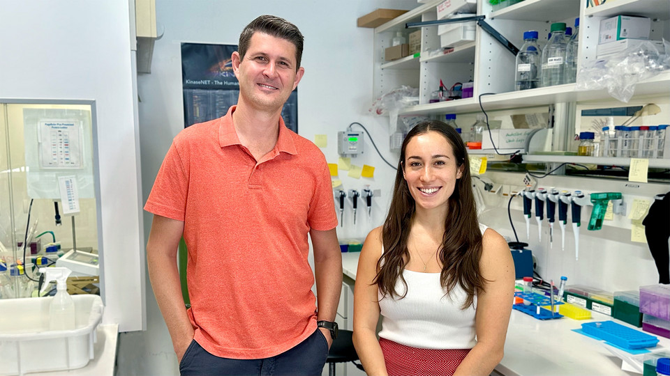
Group leader Thomas Leonard together with first author Sumire Antonioli. © Max Perutz Labs
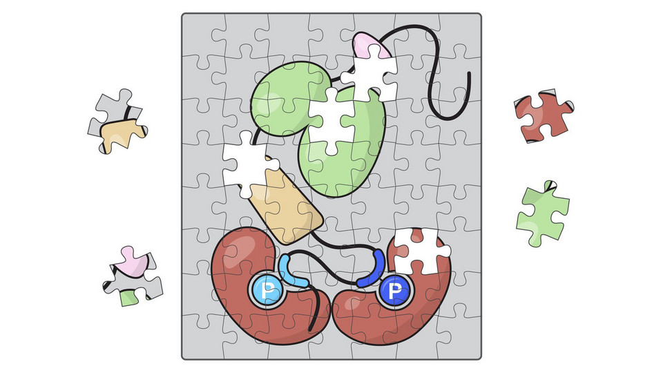
Putting the pieces together: we identify 14-3-3 eta, group II PAKs, and Tau as parts of the MAST1 signaling pathway, pointing to a link between the actin and microtubule cytoskeletons in neurodevelopment. © Sumire Antonioli
Mega corpus callosum syndrome (MCC) is a rare neurodevelopmental disorder characterized by abnormalities in brain development, including an unusually enlarged corpus callosum, the thick bundle of nerve fibers that connects the brain’s left and right hemispheres and enables communication between them. Genetic studies have linked the condition to mutations in MAST1, a member of the microtubule-associated serine/threonine kinase family. While these findings established a clear association between MAST1 and MCC, the molecular role of MAST1 in neurons has remained poorly understood. In particular, nothing is known about how the kinase is regulated, which proteins it interacts with, and how disease-associated mutations disrupt its function. A new study by the Leonard lab now begins to fill these gaps by identifying key regulators of MAST1 signaling and characterizing the impact of disease-associated mutations on MAST1 structure and activity.
To understand where MAST1 fits within neuronal signaling, the researchers mapped the proteins that interact with and regulate the kinase. They identified 14-3-3η as a binding partner of MAST1 and found that group II PAK kinases phosphorylate one of the 14-3-3η binding sites in MAST1, while the microtubule-associated protein Tau emerged as a candidate downstream substrate. Together, these interactions place MAST1 in a signaling cascade that connects PAK-dependent regulation of the actin cytoskeleton with Tau-mediated control of microtubules. “Our findings suggest that MAST1 could sit at this interface,” says first author Sumire Antonioli, pointing to a possible mechanism for coordinating two cytoskeletal systems that are essential for neuronal development. The proposed pathway lays the molecular groundwork for understanding how disrupted MAST1 signaling may alter cytoskeletal dynamics and contribute to the disease-related features and clinical manifestations of MCC.
By linking disease-associated MAST1 mutations to changes in protein folding and kinase activity, the study provides a mechanistic bridge between genetic alterations in MAST1 and the cellular defects that may contribute to MCC. Investigation of the MAST kinases is particularly challenging because they are large proteins, contain extensive intrinsically disordered regions and their expression is restricted, predominantly, to the brain. Addressing these challenges required a collaborative approach, with the Perutz Mass Spectrometry Facility playing a central role in identifying MAST1 interaction partners and phosphorylation sites. Researchers at the Ludwig Maximillian University (LMU), Munich, Germany, and the neighboring Institute of Molecular Pathology (IMP) contributed to the study by generating and analyzing a mouse model of a patient-derived MCC mutation in MAST1. “Our work adds new regulatory nodes, interaction partners, and a downstream substrate to a signaling pathway that, until now, has not received much attention, but which is increasingly being linked to a spectrum of neurodevelopmental disorders”, says group leader Thomas Leonard.
Read the study
DOI: 10.1073/pnas.2617534123