The RNA exosome is one of the cell's critical RNA degradation machines, responsible for the turnover of a wide range of RNAs. While its functions have been studied extensively for decades, a fundamental question remained unanswered: how does the 12-subunit complex precisely assemble inside living cells? For the Ameres lab, the answer emerged serendipitously while investigating RNA degradation and searching for the best way to experimentally disrupt exosome function. As first author Tsimafei Navalayeu recalls: “It started as a side project: We became curious about how the complex assembles, and what began as an exploratory experiment ultimately turned into the central story of the study.”
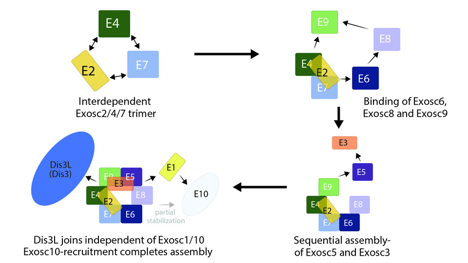
To tackle the question, the researchers established an inducible dual-guide CRISPR knockout system in mouse embryonic stem cells, enabling the controlled depletion of individual exosome subunits. By systematically removing each component, they uncovered a distinctive pattern of co-destabilization that allowed them to reconstruct the assembly pathway of the complex. Their data revealed that assembly begins with a stable Exosc2–Exosc4–Exosc7 trimer, followed by the sequential addition of the remaining subunits, demonstrating that exosome formation is a highly ordered process. “It's not just randomly 12 proteins come together and bind in whatever order. It's very specific and very tightly regulated, and this was entirely unknown,” says Tsimafei. The study therefore provides the first in vivo assembly map of the mammalian RNA exosome and reveals how its modular organization contributes to function.
Remarkably, unlike other structural subunits, the terminally incorporated cap subunit Exosc1 proved dispensable for cell viability, revealing that the exosome possesses a modular and functionally resilient architecture. The team further showed that ‘orphaned’ exosome subunits – proteins that fail to assemble in the complex – are selectively degraded by the ubiquitin–proteasome system, linking exosome assembly to cellular protein quality-control mechanisms.
For Tsimafei, one of the most memorable moments came when the emerging data suddenly made sense. “It was a very special moment when you realize that you have understood something that nobody else understands and nobody else has seen before,” he says. The insights have transformed an initial exploratory experiment into a new framework for understanding how one of the cell's most important molecular machines is built. The work was carried out entirely at the Vienna BioCenter, in collaboration with the Zuber and Plaschka labs of the Research Institute of Molecular Pathology (IMP).
DOI: 10.1038/s44318-026-00824-x