The human DNA is almost 2 meters long when unfolded, yet it is packed into microscopically visible chromosomes, which fit into a cell’s nucleus. Nature solves this fascinating packing problem by wrapping DNA around histones to create a chromatin fibre. This fibre in turn can fold into chromosomes, which become visible during mitosis.
The assembly of mitotic chromosomes is essential for the correct inheritance of our genetic material and errors in this process can lead to mis-segregation of chromosomes and human disease. The molecular mechanisms controlling the material properties and integrity of chromosomes during mitosis remain poorly understood.
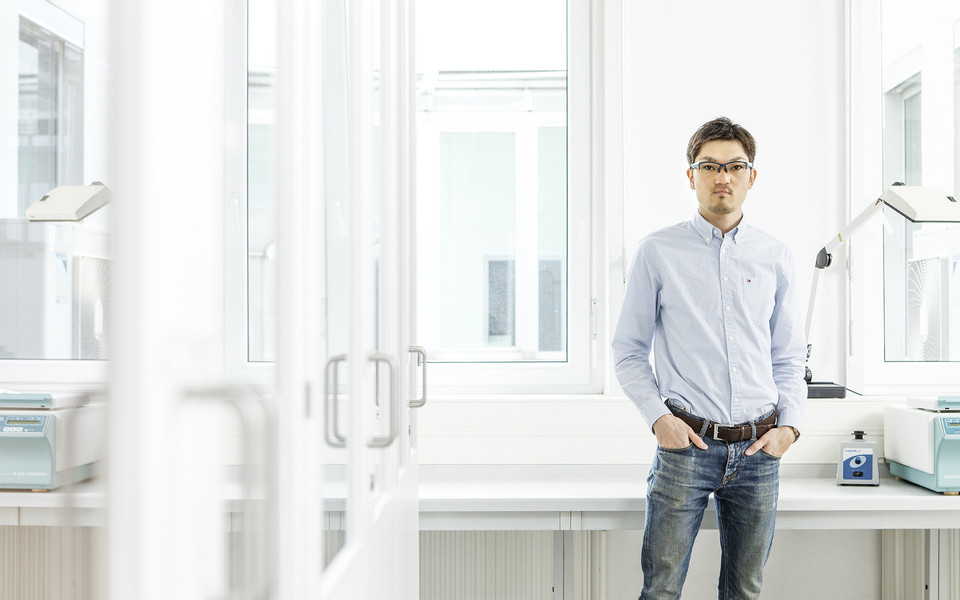
“Understanding the structural organization of chromosomes has remained a challenging goal, owing to the irregular fold of the chromatin fibre at the nanometer-scale. A complete understanding of chromosomes requires their visualization at different length scales. This can only be achieved by combining different modes of imaging. Moreover, we will investigate how epigenetic marks on histones influence chromosome folding”, says co-Principal Investigator Shotaro Otsuka.
Shotaro is an expert in advanced microscopy, and has developed a novel imaging approach that allows to observe cells simultaneously by both light and electron microscopy thereby pushing the limits of resolution in live cells. Shotaro will use such techniques to visualize chromatin during mitosis with nanometre precision.