When light passes through turbulent or dense material, such as millimetre-thick tissue, standard imaging technologies experience significant limitations in the imaging quality. Scientists therefore place deformable mirrors in the optical path of the telescope or microscope, which cancel out the undesired effects. This so-called adaptive optics has led to many breakthroughs deep-tissue imaging. However, this level of control has not yet been achieved in electron optics even though many applications in materials science and structural biology demand it. In electron optics, scientists use beams of electrons instead of light to image structures with atomic resolution. Usually, static electromagnetic fields are used to steer and focus the electron beams.
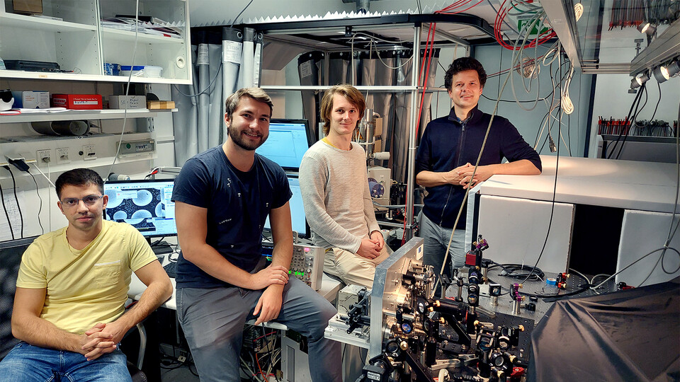
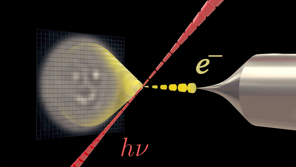
In their new study, the Juffmann lab (Faculty of Physics and the Max Perutz Labs) and their colleagues from the University of Siegen now show that it is possible to deflect electron beams almost arbitrarily using high-intensity, shaped light fields, which repel electrons. The scientists’ experiment makes use of our ability to shape light. A laser pulse is shaped by a spatial light modulator and interacts with a counter-propagating, synchronized pulsed electron beam in a modified scanning electron microscope. This enables imprinting, on demand, transverse phase shifts to the electron wave, enabling unprecedented control over electron beams.
“We are writing with the laser beam in the transverse phase of the electron wave. Our experiments pave the way for wavefront shaping in pulsed electron microscopes with thousands of programmable pixels. In the future, parts of your electron microscope may be made from light,” says lead author of the study, Marius Mihaila Constantin Chririta. In contrast to other competing electron-shaping technologies, the scheme is programmable, and avoids losses, inelastic scattering, and instabilities due to the degradation of material diffraction elements. Group leader Thomas Juffmann concludes: “Our shaping technique enables aberration correction and adaptive imaging in pulsed electron microscopes. It can be used to adjust microscopes to the studied specimens to maximize sensitivity.”
Publication:
Marius Constantin Chirita Mihaila, Philipp Weber, Matthias Schneller, Lucas Grandits, Stefan Nimmrichter, and Thomas Juffmann: Transverse Electron-Beam Shaping with Light. Physical Review X. 2022.