

First author Elisabeth Holzer together with group leader Sascha Martens. © Max Perutz Labs
All authors - from left to right: Martina Schuschnig, Julia Romanov, Elisabeth Holzer, Justyna Sawa-Makarska, and Sascha Martens. © Martens lab
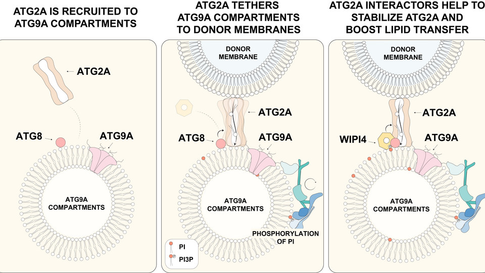
Our findings reveal how ATG9 compartments recruit the lipid transfer protein ATG2, which transfers, among other lipids, phosphatidylinositol (PI) to these compartments, providing a source for PI3P production that supports the downstream regulation of autophagosome formation. © Elisabeth Holzer
Autophagy is a fundamental cellular recycling pathway that degrades damaged, excess, or potentially harmful cellular material. Much of what we know about this process comes from studies in yeast, a genetically tractable eukaryotic model system that has been instrumental in identifying the core machinery of autophagy. During autophagosome biogenesis, a small precursor membrane expands into a cup-shaped phagophore, which gradually encloses its cargo before closing to form a mature autophagosome. Although many components of the autophagy machinery are conserved from yeast to mammals, the mechanisms that regulate this membrane-remodeling process exhibit some significant differences. A new study from the Martens lab highlights how essential lipids are delivered to developing autophagosomes, thereby providing insight into the earliest steps of their formation in mammalian cells.
At the center of the study is ATG9A, a lipid scramblase and the only transmembrane protein of the core autophagy machinery. Lipidomic analysis revealed that mammalian ATG9A-containing compartments contain only minimal levels of phosphatidylinositol (PI) compared to their yeast counterparts. The researchers showed that the lipid-transfer protein ATG2A delivers PI to these precursor membranes, a step that is essential for autophagosome formation. First author Elisabeth Holzer, a PhD student in the Martens lab, explains: “Without ATG2 there is no autophagy at all, because no lipids can be transported into the seed membrane and therefore the membrane cannot expand.” The study additionally identified ATG8-like proteins as key factors that recruit and activate ATG2A to drive membrane growth.
“The precursor membranes are the seeds for an organelle that can form within 10 minutes. They are very small at the beginning but then grow into super-large compartments that can engulf even a bacterium or whole organelles,” Elisabeth says. The findings support a model in which a positive feedback loop involving ATG2A, ATG8 proteins and WIPI4 activates ATG9A-containing precursor membranes for phagophore expansion. PI delivered by ATG2A is converted by a lipid kinase, PI3KC3, into PI3P, which recruits WIPI4 and, in turn, additional ATG2A, thereby progressively amplifying lipid transport to the growing membrane. “At the beginning, we think that the lipid transport is slow, but that this feedback loop facilitates membrane expansion into a full autophagosome,” Elisabeth explains.
Understanding how autophagy works – and why it sometimes fails – is particularly important in long-lived cells such as neurons. When damaged cellular material is not efficiently removed, normal cellular physiology is adversely affected, contributing, ultimately, to cell death, a hallmark of neurodegenerative diseases such as Parkinson’s Disease. Since most neurons do not readily regenerate, their loss can have lasting consequences. By uncovering fundamental mechanisms that control autophagosome formation, the study helps build the molecular framework needed to understand how defects in this pathway contribute to neurodegeneration. The work was supported by the Aligning Science Across Parkinson’s (ASAP) initiative, in partnership with The Michael J. Fox Foundation for Parkinson’s Research (MJFF).
DOI: 10.1038/s41467-026-77368-5